
Efferocytosis And Its Oft-Unstated Role In Artery Clogging.
There is a natural biological mechanism in our bodies that prevents the arterial plaque formation. The only problem is how we can keep it running smoothly.

The word “efferocytosis” isn’t one that is frequently thrown about these days. Most heart disease patients would not have heard of this word before, because rarely is it (if ever) discussed by their doctors.
Oddly, though, it is a biological process that occurs naturally within our bodies. Much like autophagy, which more people would have heard of:
Autophagy - How Our Body Performs a Self-Renewing Process

Our bodies consist of approximately 38 trillion cells. These cells are present in just about every part of our body as parts of various different systems and carry out various different functions to support the health of the human body as a whole. The human body, therefore, can be likened to an organisational structure. In the grand scheme of things, an …
Of course, before dead cells are destroyed, they have to be programmed to commit suicide first. There’s a marked difference when we have dead skin that sloughs off as compared to flaps of dead skin that we have to cut off when we suffer an injury. Somehow, those flaps of skin still can cause pain, while skin that sloughs off is painless.
Because programmed suicide is easier to deal with than a sudden, accidental death.
It happens in real life too - it’s more about how much control one has over their own fates.
In the realm of programmed suicides, a cell that is programmed to commit suicide undergoes apoptosis.
It is stated in this article that “the human body turns over more than one million cells per second through a process known as programmed cell death (PrCD), or apoptosis”.
That article goes on further to state that “the body engages in an evolutionarily conserved process known as programmed cell removal (PrCR), or efferocytosis” to remove those dead cells.
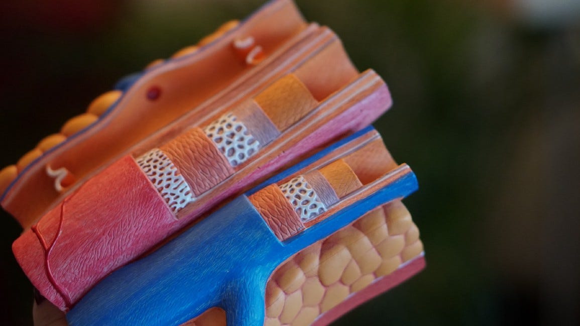
And we can see this process occurring in atherosclerotic plaques, too.
Now, Seriously, What's So Tricky About Cholesterol?

This article is the final part of a 3 part miniseries. The first 2 parts can be found at: Medical science has been talking about cholesterol and its links to heart disease for years. People who are at risk of heart failure are invariably prescribed medicines such as
These plaques contain a necrotic core that is rich in dead apoptotic cells. If all these dead cells aren’t cleared in time, they will contribute to the development of a pro-inflammatory environment.
The pro-inflammatory environment significantly affects the cells that are requisite for conducting efferocytosis, which happens to be the macrophage cells in the body’s immune system.
They are generally present as 2 different phenotypes: the M1 and the M2 variants.
This article suggests that a macrophage is initially neutral, but it polarises into the M1 or M2 phenotype based on the prevailing cytokine signal. The pro-inflammatory cytokine tumour necrosis factor alpha (TNF-α), for instance, promotes M1 polarisation.
And it is also known that “increased concentrations of TNF-α are found in acute and chronic inflammatory conditions”, as reported in this article.
So when we experience an injury, for instance, we will experience a bout of acute inflammation around the injured region - this inflammation will trigger a preferential differentiation of new macrophage cells into the M1 variant to conduct phagocytosis on the injured/dead muscle cells and clear out cellular debris.
When their job is done, they hand over the next set of duties for the M2 macrophages to conduct, and that is when the inflammatory signal intensity will subside. When the M1 guys have not completed their end of the work, the inflammation signal ought to remain, and with it any associated symptoms of pain, aches and/or swelling.
But let’s get back to the topic of heart disease.
The M2 macrophages are also responsible for conducting efferocytosis.
However, let’s not forget that the amount of dead cells in the atherosclerotic plaques lining the arteries of heart disease patients can be significant. Significant to the point that they’re pumping in sufficient quantities of pro-inflammatory cytokines to force the differentiation of new neutral macrophages primarily into the M1 phenotype.
The question then becomes “where are the necessary M2 macrophages to conduct efferocytosis on the plaques, then?” The simple answer is that the M2 population would end up getting smaller over time with chronically elevated inflammatory signals as every new macrophage preferentially differentiates into the M1 phenotype.
If most of the macrophages are of the M1 variant, we’d see much less M2s being able to conduct efferocytosis - and if the rate of efferocytosis is impaired, good luck seeing a decrease in the size of the atherosclerotic plaques in an artery.
Therefore, can atherosclerotic plaque sizes be reduced?
Yes, they can - BUT we have a whole mountain of work ahead of us when dealing with the chronic inflammation that an atherosclerotic patient is doubtless facing.
And then it becomes a race against time - can we change the inflammatory environment in a patient’s body significantly enough to improve the rate of efferocytosis before the plaque ruptures to trigger a heart attack, which, by the way, is related to the activity of live foam cells that are entrapped within the plaque (and is not linked to the efferocytosis activity)?
If we can - the plaques will eventually shrink over time.
If we can’t - no amount of stents, bypasses or heart surgeries will prevent the clogging of arteries, really.
The Uncomfortable Truth Behind Healthcare Strategies Targeting Heart Disease

According to the World Health Organization, heart disease is the number one killer of humans worldwide. And as such, there have been many different things that the healthcare industry has developed with in an attempt to counteract that issue, such as:
Which would explain how some people can change up their lifestyles significantly enough to experience an overall reduction in their plaque sizes, while others end up dying from heart attacks before they can even get to plaque reduction.
It’s a race against time when the doctor diagnoses one with clogged arteries - that’s a major indicator that efferocytosis isn’t working properly in the person’s body already.
And if efferocytosis wasn’t functioning efficiently enough, what can we say about that person’s ability to heal from injuries or to recover from illnesses?
Those recovery rates would be markedly lower too, wouldn’t it?
It’s an immunological issue at the heart of it all!
If you were to refer to the nutrients that support immune system functions or heart health, you’d find out that there is a ton of nutrients in common. And that’s because heart health is heavily dependent on the immune system:
10 Nutrients That Support A Healthy Heart

Supporting cardiovascular health is not an easy thing to do. We eat food. We exercise. We get stressed by work. We sleep. All these actions do contribute to the overall state of our immune system. As a result, if we ever do encounter situations such as high blood cholesterol in our middle age years (or later), we freeze. We don’t really know what to do. …
Nutrients That Support A Healthy Immune System

In Making Sense Of Our Immune System, I do highlight the main problems that our immune system faces as its functionality declines based on poor sustained lifestyle habits that we are subscribing to. In that article, I also do highlight what aspects of can go wrong with our immune systems without us realising it, even as we seek to support our immune syst…
We can’t really disconnect our heart from our immune system!
Do feel free to share this article and hit the “subscribe” button to get more updates about the science concepts in nutrition and health, all deconstructed nicely for your convenient perusal!
Doc: What about extracting a person's own M2 macrophages, multiplying them and then infusing them back into the blood? That's simplistic, I know, because of the signaling issues involved. Or if we know exactly how the M2 macrophages reduce plaques, perhaps create bio-nanoparticles that do the same thing but can't be switched off; once they home in on a plaque, they just dissolve it or eat it away until it dissolves? I know there would be the risk of it breaking loose at a certain point but there's that now, and leaving it alone eventually wreaks havoc anyway. This is an engineering and SIGINT problem. Hard to believe creative solutions haven't been found.